
Iron, Dopamine & PMDD: Connecting the Dots

If you have PMDD, you have probably been told it is a hormone problem. That your estrogen and progesterone are fluctuating, that your brain is sensitive to those fluctuations, and that the options are antidepressants, hormonal birth control, or learning to manage.
What you have likely not been told is that iron status may be quietly shaping the severity of what you experience every luteal phase, through its direct role in dopamine production, and through the way dopamine and estrogen are biochemically intertwined across the menstrual cycle.
This is not a simple story. The research connecting iron specifically to PMDD is sparse, and that gap itself is worth naming. But the mechanistic pathway is coherent, the adjacent evidence is meaningful, and in clinical practice, this pattern shows up consistently enough that it deserves a clear explanation.
PMDD Is Not a Hormone Level Problem
One of the most important shifts in how PMDD is understood is the recognition that women with PMDD generally have normal estrogen and progesterone levels. What differs is how the brain and nervous system respond to the normal fluctuations of those hormones across the cycle.
Research consistently shows that PMDD reflects altered central nervous system sensitivity to neuroactive steroid hormones, not abnormal hormone quantities (Hantsoo & Epperson, 2020). The diagnosis does not come with a hormonal biomarker. It comes with a pattern: symptoms that emerge reliably in the luteal phase and resolve shortly after menstruation begins.
Understanding why requires looking at what is happening at the neurotransmitter level and specifically, at dopamine.

Estradiol as a Dopamine Amplifier
Estradiol (the most potent form of estrogen) is not just a reproductive hormone. It is a significant modulator of dopaminergic signaling throughout the brain, and its fluctuations across the menstrual cycle directly shape mood, motivation, reward sensitivity, and emotional resilience.
Estradiol works on multiple nodes of the dopamine system simultaneously. It stimulates dopamine synthesis by activating tyrosine hydroxylase, the rate-limiting enzyme in dopamine production. It enhances dopamine release in the nucleus accumbens, the brain's primary reward center. And it slows dopamine reuptake, keeping more dopamine available in the synapse for longer.

A landmark 2025 study published in Nature Neuroscience demonstrated this mechanism directly. Golden and colleagues measured dopamine reward prediction errors in the nucleus accumbens across the reproductive cycle and found that endogenous rises in 17β-estradiol predicted significantly larger dopamine reward signals and greater behavioral sensitivity to rewards. Proteomics analysis revealed that estradiol increases were accompanied by reduced dopamine transporter expression, the molecular mechanism by which estradiol slows reuptake and keeps dopamine available (Golden et al., 2025).
An earlier neuroimaging study by Dreher and colleagues found that women showed greater activation of the midbrain, striatum, and orbitofrontal cortex during reward delivery in the midfollicular phase (when estradiol is rising) compared to the luteal phase (Dreher et al., 2007). High estradiol means enhanced dopaminergic reward processing. The brain is more motivated, more responsive, more regulated.
And then estradiol drops.
The Luteal Phase as Dopamine Withdrawal
In the late luteal phase (the one to two weeks before menstruation) estradiol falls. Progesterone, which rose after ovulation, also begins to decline. The brain loses its primary dopaminergic amplifier at exactly the moment it is also navigating the fluctuations of allopregnanolone and GABA receptor sensitivity.
Research has framed this premenstrual estrogen withdrawal as functionally analogous to dopamine withdrawal. Ossewaarde and colleagues used fMRI during a monetary incentive task and found enhanced ventral striatal responses to reward cues in the premenstrual phase compared to the follicular phase, a pattern they explicitly described as consistent with a dopamine withdrawal state, where heightened reward-cue reactivity signals the brain compensating for reduced dopaminergic tone (Ossewaarde et al., 2010). This compensatory response was strongest in women with more premenstrual symptoms.
A review by Yoest, Quigley, and Becker synthesized the broader literature, noting that estradiol rapidly enhances dopamine release in both the dorsal striatum and nucleus accumbens, and that both estradiol and progesterone show time-dependent patterns of initial enhancement followed by later inhibition, contributing to the characteristic instability of the late luteal phase (Yoest et al., 2018).
A randomized controlled trial by Schmidt and colleagues provided perhaps the most direct clinical evidence. Postmenopausal women with a history of perimenopausal depression who were withdrawn from estradiol under blinded conditions experienced significant recurrence of depressive symptoms (their depression scores roughly tripling) while those who continued estradiol remained asymptomatic. The researchers concluded that it is the change in ovarian steroids, not sustained levels above a threshold, that precipitates mood symptoms (Schmidt et al., 2015).
For women with PMDD, this monthly estradiol withdrawal is not a minor fluctuation. It is a predictable neurochemical event that disrupts the dopaminergic system the brain has come to rely on across the follicular phase.

The Allopregnanolone Layer
Dopamine is not the only system destabilized in the luteal phase. Allopregnanolone, a progesterone metabolite and potent positive modulator of the GABA-A receptor, follows a parallel trajectory, rising across the luteal phase and dropping sharply in the days before menstruation.
In most women, allopregnanolone has a calming, sedating effect. It potentiates GABA (the brain's primary inhibitory neurotransmitter) reducing anxiety, attenuating the stress response, and helping regulate HPA axis activity. In women with PMDD, this system does not work the way it should.
The critical insight from Hantsoo and Epperson's (2020) comprehensive review is that women with PMDD have normal allopregnanolone levels, but dysregulated GABA-A receptor sensitivity to those fluctuations. The receptor fails to adapt appropriately to the rise and fall of allopregnanolone across the cycle. When allopregnanolone drops sharply in the late luteal phase, the GABA-A receptor upregulates its α4 subunit dramatically — a structural change associated with increased anxiety and irritability rather than calm.
A randomized controlled trial by Martinez and colleagues tested this mechanism directly. Using dutasteride (a drug that blocks the enzyme converting progesterone to allopregnanolone) researchers stabilized allopregnanolone levels in women with PMDD while allowing normal progesterone fluctuations. Stabilizing the rate of ALLO change, rather than the absolute level, reduced symptoms of irritability, low mood, anxiety, food cravings, and bloating. This confirmed that it is the fluctuation in allopregnanolone, not the level itself, that triggers PMDD symptoms in susceptible individuals (Martinez et al., 2016, as cited in Hantsoo & Epperson, 2020).
Because allopregnanolone normally attenuates cortisol release by potentiating GABAergic transmission, its dysregulated receptor sensitivity also impairs HPA axis regulation. This is why stress sensitivity is specifically heightened in the luteal phase in PMDD — the system that is supposed to buffer stress is not functioning normally during the exact window when it is most needed (Hantsoo & Epperson, 2020).
So in the late luteal phase, a woman with PMDD is navigating simultaneous dopamine withdrawal from falling estradiol and dysregulated GABA buffering from allopregnanolone fluctuations. Two systems failing to buffer each other at the same time.
This is where iron enters the picture.

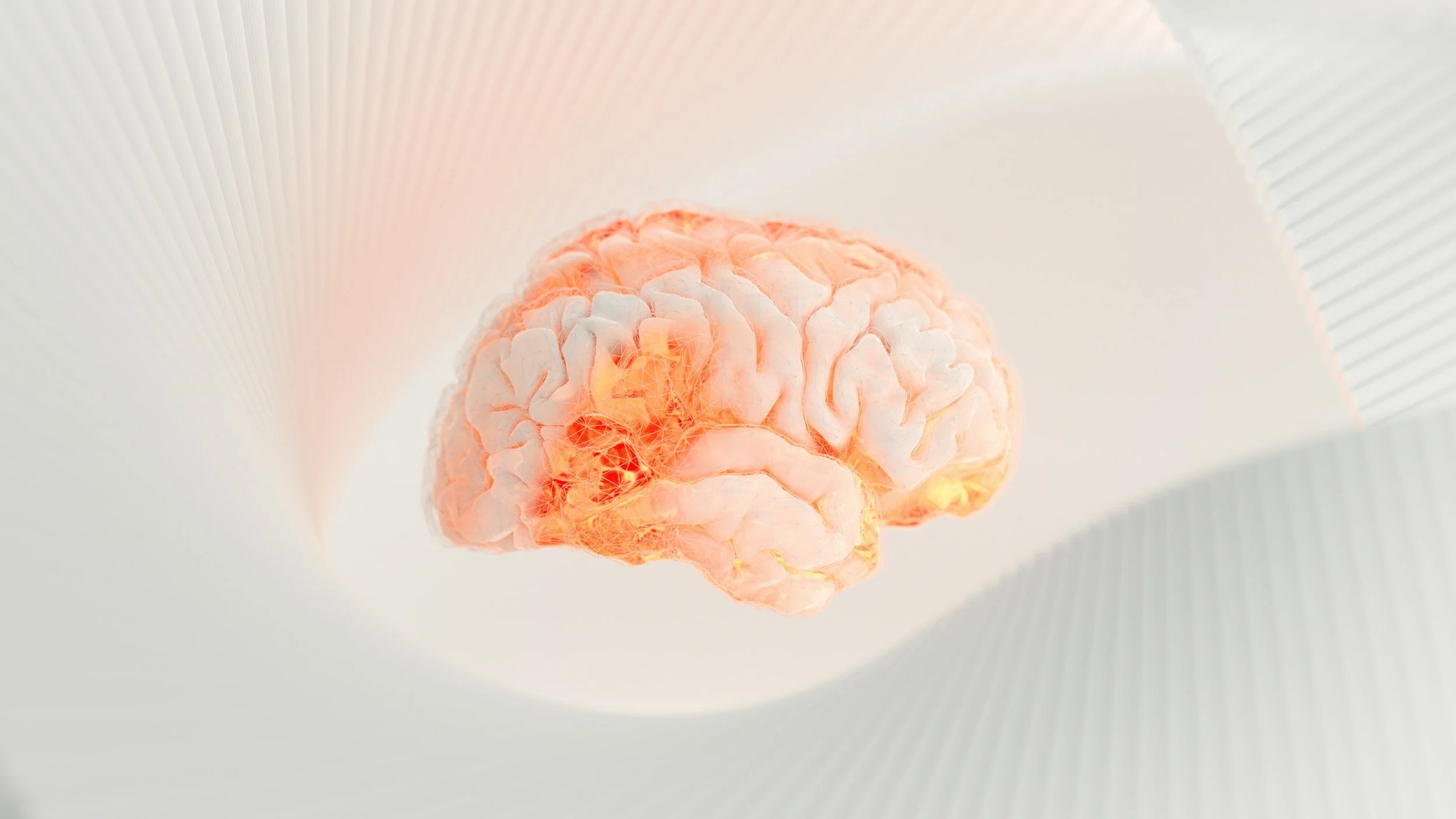
Iron and Dopamine: The Missing Piece
Iron is an essential cofactor for tyrosine hydroxylase — the rate-limiting enzyme in dopamine synthesis. Without adequate iron, the brain cannot produce dopamine efficiently. When iron stores are low, dopamine production slows, dopamine receptor density decreases, and dopamine signaling is impaired in the striatum and prefrontal cortex — the same brain regions most affected by the luteal estradiol withdrawal described above (Hare & Double, 2016; Lozoff, 2020).
Research has consistently demonstrated that low ferritin is associated with more severe symptoms in conditions driven by dopamine dysregulation. Low ferritin is consistently associated with more severe ADHD symptoms, and iron deficiency reduces dopamine receptor density and disrupts dopaminergic signaling in the striatum and prefrontal cortex through mechanisms directly relevant to mood and emotional regulation (Nowak et al., 2021; Gustavsson et al., 2023).

The iron-dopamine relationship also runs in both directions. When iron is too low, dopamine production stalls. When unbound iron is too high, oxidative stress can damage dopaminergic neurons. Hare and Double described this as a toxic couple — both essential and potentially harmful when unbalanced (Hare & Double, 2016).
Now consider what this means for the luteal phase. Estradiol is falling, removing its dopamine-amplifying effects. Allopregnanolone fluctuations are destabilizing GABA buffering. And if iron stores are also insufficient, the brain's capacity to synthesize dopamine in the first place is compromised. The deficit compounds.
Does Iron Status Affect PMDD Directly?
The direct research connecting iron specifically to PMDD is sparse and that honesty matters here. There is currently no study that has measured ferritin across the menstrual cycle in women with diagnosed PMDD, no intervention trial using iron supplementation for PMDD, and no study correlating luteal-phase iron fluctuations with PMDD symptom severity.
What exists is adjacent and suggestive.
A small cross-sectional study of women with PMDD found significantly lower hemoglobin in the PMDD group compared to controls (11.3 versus 12.6 g/dL) along with elevated inflammatory markers (Khan et al., 2023). A Mendelian randomization study by Zeitoun and colleagues found that women with genetically elevated risk of iron overload were less likely to report premenstrual symptoms including confusion, headaches, and nausea, pointing toward a protective direction for iron sufficiency, even if the reverse (low iron worsening symptoms) was not clearly demonstrated in that sample (Zeitoun et al., 2021).
A review by Rajkumar noted that the iron-depression link appears strongest during specific life stages characterized by both high iron demand and significant hormonal flux, adolescence, late pregnancy, and postpartum, framing the mechanistic connection between iron, monoamine synthesis, and mood as most clinically relevant during hormonal transitions (Rajkumar, 2026).
The bridge between iron insufficiency and PMDD severity is mechanistic rather than empirically established. But the mechanism is coherent, the adjacent evidence is consistent with it, and the clinical picture is familiar to anyone working in this space.

Why Menstruating Women Are Especially Vulnerable
Iron insufficiency is not evenly distributed across the population. Premenopausal women carry a fundamentally different iron burden than men or postmenopausal women — because they lose iron every month.
Large Danish population surveys found median ferritin of 37 to 42 µg/L in premenopausal women compared to 71 to 80 µg/L in postmenopausal women, with approximately 18% of premenopausal women having depleted iron stores and another 23% having marginal stores, meaning roughly 40% of premenopausal women are operating with suboptimal ferritin (Milman et al., as cited in research summary). For women with heavy menstrual bleeding, the picture is significantly worse. Napolitano and colleagues measured actual menstrual iron losses and found that women with menorrhagia lost approximately 5.2 mg of iron per cycle, five to six times more than the roughly 1 mg lost in typical cycles, with mean ferritin of just 6.4 ng/mL compared to 36.2 ng/mL in controls (Napolitano et al.).

Among adolescents with heavy menstrual bleeding, studies have found that 61 to 87% have ferritin at or below 40 ng/mL, with the majority reporting significant fatigue, a pattern that frequently goes undetected because hemoglobin and CBC testing miss iron deficiency without anemia in more than half of affected patients (Zia et al.; Wang et al.; Lang et al.).
The conventional ferritin cutoff of 15 µg/L (still used by many laboratories) increasingly appears inadequate for identifying functional iron deficiency. A pooled analysis of stable iron isotope studies in over 1,000 women found that the body begins upregulating iron absorption when ferritin falls below approximately 50 µg/L, suggesting that incipient iron deficiency begins well above the traditional threshold (Galetti et al., 2021). An analysis of ROC data found an optimal ferritin cutoff of 45 µg/L for females to diagnose iron deficiency, improving diagnostic sensitivity by 44% over current reference ranges (Troike & McShane, 2025).
In clinical practice, this means a woman can receive a "normal" ferritin result and still be operating with iron stores too low to support optimal dopamine synthesis — particularly in the luteal phase, when dopamine demand is already under pressure from falling estradiol.
How These Pieces Connect
Putting this together in clinical terms:
A woman with PMDD enters the luteal phase with falling estradiol removing the brain's primary dopaminergic amplifier. Allopregnanolone fluctuations are destabilizing GABA-A receptor sensitivity, impairing stress buffering. And if her ferritin is sitting at 18 µg/L (technically within the conventional normal range) her brain is also starting the dopamine synthesis process at a disadvantage, because tyrosine hydroxylase requires adequate iron to function optimally.
Each of these is a partial impairment. Together, they compound. The result is not just mood instability. It is mood instability with a physiological explanation — one that conventional assessments rarely capture because the pieces are evaluated separately rather than as an interacting system.
This is not a reason for despair. It is a roadmap.
What Functional Assessment Can Reveal
Standard care for PMDD rarely includes ferritin testing and when it does, the threshold used may not reflect functional iron sufficiency for brain health. Functional medicine takes a broader view.

In working with clients who have PMDD, I look at ferritin with optimal brain function ranges in mind (typically 50 to 90 ng/mL) not just conventional normal ranges. I assess the full iron panel including transferrin saturation and TIBC to understand whether iron is low due to insufficient intake, poor absorption, or chronic inflammation. The GI-MAP can reveal gut dysbiosis, inflammation, or pathogens that impair iron absorption even when dietary intake appears adequate. The DUTCH test shows progesterone metabolism, cortisol patterns, and estrogen clearance, providing a full hormonal picture alongside the nutritional one. The Organic Acids Test reveals downstream markers of neurotransmitter metabolism and oxidative stress that reflect how the dopamine system is actually functioning.
These pieces together, iron status, gut health, hormonal metabolism, neurotransmitter patterns — give a much clearer picture of what is driving symptom severity in any individual woman than a single hormone panel ever could.
PMDD is not simply a hormone disorder. For many women, it involves a convergence of neurobiological sensitivity, dysregulated GABA-A receptor function, falling estradiol removing dopaminergic support, and (potentially) insufficient iron stores that compromise the brain's capacity to synthesize dopamine at exactly the wrong moment in the cycle.
The research connecting iron directly to PMDD is still emerging. But the mechanistic pathway is coherent, the adjacent evidence is consistent, and the clinical pattern is recognizable. Evaluating iron status as part of PMDD assessment is not a stretch. It is a logical extension of understanding what the brain needs to regulate dopamine and what menstruating women are uniquely at risk of losing every month.
If this connects dots you have been trying to connect on your own, that recognition is worth paying attention to.
PMDD rarely exists in isolation. When I work with clients navigating PMDD, I look at the full picture — hormonal metabolism, iron and nutrient status, gut health, stress physiology, and neurotransmitter patterns. Because the luteal phase does not just reveal hormonal sensitivity. It reveals everything the nervous system is trying to compensate for.
Let’s talk through your pattern and whether functional lab testing makes sense for you.
References
Dreher, J. C., Schmidt, P. J., Kohn, P., Furman, D., Rubinow, D., & Berman, K. F. (2007). Menstrual cycle phase modulates reward-related neural function in women. Proceedings of the National Academy of Sciences, 104(7), 2465–2470. https://doi.org/10.1073/pnas.0605569104
Galetti, V., Stoffel, N. U., Sieber, C., Zeder, C., Moretti, D., & Zimmermann, M. B. (2021). Threshold ferritin and hepcidin concentrations indicating early iron deficiency in young women based on upregulation of iron absorption. EClinicalMedicine, 39, 101052. https://doi.org/10.1016/j.eclinm.2021.101052
Golden, C. E. M., Martin, A. C., Kaur, D., Mah, A., Levy, D. H., Yamaguchi, T., Lasek, A. W., Lin, D., Aoki, C., & Constantinople, C. M. (2025). Estrogen modulates reward prediction errors and reinforcement learning. Nature Neuroscience, 28, 2502–2514. https://doi.org/10.1038/s41593-025-02104-z
Gustavsson, J., Johansson, J., Falahati, F., Andersson, M., Papenberg, G., Avelar-Pereira, B., Bäckman, L., Kalpouzos, G., & Salami, A. (2023). The iron-dopamine D1 coupling modulates neural signatures of working memory across adult lifespan. NeuroImage, 279, 120323. https://doi.org/10.1016/j.neuroimage.2023.120323
Hantsoo, L., & Epperson, C. N. (2020). Allopregnanolone in premenstrual dysphoric disorder (PMDD): Evidence for dysregulated sensitivity to GABA-A receptor modulating neuroactive steroids across the menstrual cycle. Neurobiology of Stress, 12, 100213. https://doi.org/10.1016/j.ynstr.2020.100213
Hare, D. J., & Double, K. L. (2016). Iron and dopamine: a toxic couple. Brain : a journal of neurology, 139(Pt 4), 1026–1035. https://doi.org/10.1093/brain/aww022