
Your Labs Came Back Normal. Your Symptoms Didn't Get the Memo.

You have heard it before. Maybe more times than you can count.
Everything looks fine.
Your doctor reviewed the results, told you your numbers are within range, and sent you home. And yet you are still exhausted in a way that sleep does not fix. Your digestion is unpredictable. Your mood crashes without warning. You cannot lose weight no matter what you do. You keep getting sick. Something is clearly wrong, and yet every test says otherwise.
This is not a mystery. And it is not in your head.
It is a gap between what standard lab work is designed to find and what is actually driving your symptoms. Those are not the same thing. And understanding the difference is often where the answers finally start to appear.
What Standard Blood Work Is Actually Designed to Do
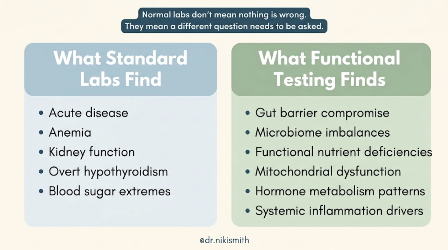
Standard lab panels, including the comprehensive metabolic panel, complete blood count, and basic thyroid screen, were designed with a specific purpose: to rule out acute disease. They are the right tools for detecting kidney failure, anemia, uncontrolled diabetes, and overt hypothyroidism. They are not designed to assess the functional state of your metabolism, the integrity of your gut lining, the output of your gut microbiome, or whether your mitochondria are producing energy efficiently.
That is not a flaw in the tests. It is a matter of what they were built for.
The problem is that the conditions that make people feel chronically unwell, the patterns that drive persistent fatigue, unexplained weight changes, mood instability, gut dysfunction, recurring illness, and hormonal disruption, often do not show up on those panels at all. They require a different set of questions.
The Questions Standard Testing Does Not Ask
When someone comes to me after years of being told their labs are normal, I am not starting from scratch. I am asking a different set of questions about the same body. Here is what those questions look like and why they matter.
Is your gut barrier intact?
The intestinal lining is a single cell layer thick. It is both a gateway and a gatekeeper, allowing nutrients to pass through while preventing harmful microbes, bacterial fragments, and undigested proteins from crossing into circulation. When that barrier is compromised, the downstream effects are wide-ranging and often look completely unrelated to digestion.
Elevated calprotectin, a marker of active gut inflammation released by immune cells in the gut lining, can indicate mucosal inflammation that produces no obvious GI symptoms but drives systemic inflammatory burden. Low secretory IgA, the immune system's primary defense at the gut surface, suggests compromised mucosal immunity, often seen with chronic stress, nutrient depletion, or dysbiosis. Neither appears on a standard metabolic panel (Diagnostic Solutions Laboratory, 2024).

What is your microbiome actually producing?
The gut microbiome is not a passive collection of bacteria. It is a metabolically active ecosystem whose output directly affects your immune function, hormone metabolism, neurotransmitter production, and energy regulation.
Butyrate, a short-chain fatty acid produced by specific gut bacteria including Faecalibacterium prausnitzii and Roseburia, is the primary fuel source for the cells lining your colon. It also modulates immune regulation, supports the gut barrier, and influences inflammatory signaling throughout the body. Low butyrate-producing capacity, detectable through microbiome testing, is one of the most consistent findings in people with autoimmune conditions, metabolic dysfunction, and chronic fatigue (Martino et al., 2022).
Beta-glucuronidase is an enzyme produced by specific gut bacteria that regulates how estrogen and other compounds are reabsorbed from the gut. When beta-glucuronidase activity is dysregulated, it can affect estrogen recirculation in ways that contribute to hormonal imbalance, even when standard hormone panels look normal. Interpreting this marker requires the full microbiome picture, not just the single number (Kwa et al., 2016).
Urolithin-producing bacteria convert dietary compounds from pomegranates, berries, and walnuts into urolithin A, a compound that supports mitochondrial health and cellular energy production. Whether you can actually produce urolithins from the foods you eat depends entirely on whether you have the right gut bacteria. Two people eating the same diet can have completely different physiological outcomes based on this single microbiome variable (Singh et al., 2022).
Are your B vitamins actually working at the cellular level?
Serum B12 measures how much vitamin B12 is circulating in your blood. It does not measure whether B12 is functionally available inside your cells. Methylmalonic acid (MMA), a marker on the organic acids test, accumulates when B12 is functionally insufficient, even when serum B12 looks completely normal. The distinction matters because functional B12 deficiency affects nerve function, red blood cell production, and methylation, and it will not be caught by a standard panel (Diagnostic Solutions Laboratory, 2024).
The same pattern holds for vitamin B6. Xanthurenic acid elevation on the organic acids test is one of the earliest and most sensitive functional markers of B6 insufficiency, appearing before serum B6 levels drop outside the reference range. B6 is a required cofactor for serotonin, dopamine, and GABA synthesis. Functional deficiency here affects mood, cognition, and nervous system regulation in ways that a serum B6 level would completely miss (Tada et al., 1967; Ciorba, 2013).

Are your mitochondria producing energy efficiently?
Mitochondria convert food into ATP, the energy currency your cells run on. When this process is disrupted, the result is fatigue that does not respond to rest, cognitive fog, and a persistent sense that the body is running on depleted reserves.
Krebs cycle organic acids on the OAT, including succinic acid, fumaric acid, and malic acid, reflect where the energy production process is congested and which nutrient cofactors may be insufficient. Elevated lactate and pyruvate indicate a shift toward anaerobic metabolism, often associated with thiamine insufficiency or mitochondrial stress. These patterns are clinically actionable but completely invisible on standard blood work (Shayota, 2023; Kansakar et al., 2017).
What This Looks Like in Practice
The clients I see most often have already been through the conventional system. They have had standard blood work, sometimes multiple times. They have been told everything looks fine, or that their symptoms are stress, aging, or anxiety. Many have been offered medication to manage the symptoms without any investigation into what is driving them.
When we look deeper, the picture is almost always more specific than anyone expected. Here is what functional testing commonly finds in people who were told their labs were normal:
Gut barrier compromise driving systemic inflammation and immune activation that shows up as fatigue, skin issues, joint pain, and mood instability
Low butyrate-producing capacity contributing to impaired gut integrity, reduced immune regulation, and metabolic dysfunction
Functional B12 deficiency confirmed by elevated MMA despite normal serum B12, affecting energy, cognition, and neurological function
Functional B6 insufficiency confirmed by elevated xanthurenate, affecting neurotransmitter synthesis and mood regulation
Mitochondrial dysfunction visible in Krebs cycle organic acids, driving fatigue that does not improve with sleep or rest
Dysregulated beta-glucuronidase activity affecting estrogen metabolism and contributing to hormonal instability despite normal hormone panels
Low urolithin-producing capacity limiting cellular energy support regardless of dietary choices
None of these appear on a standard comprehensive metabolic panel. All of them are real, measurable, and clinically relevant. And all of them have pathways for support when identified.
Why This Is Not About Replacing Your Doctor
Functional lab testing is not an alternative to conventional medicine. It is an additional layer of investigation that asks different questions. The goal is not to contradict what your doctor has told you, but to fill in the gaps that standard testing was not designed to find.
Many of the clients I work with are also seeing primary care physicians, specialists, and other practitioners. The functional picture often helps explain symptoms that have remained unresolved in conventional care, and can support more targeted conversations with the rest of the care team.
If your labs came back normal and you still feel terrible, that is not a reason to give up. It is a signal that the right questions have not been asked yet.
Want to Know What Is Actually Driving Your Symptoms?
A free discovery call is the best place to start. We will talk through your history, your symptoms, and what functional testing might actually show in your specific picture.
References
Ciorba, M. A. (2013). Kynurenine pathway metabolites: Relevant to vitamin B-6 deficiency and beyond. The American Journal of Clinical Nutrition, 98(4), 834–835. https://doi.org/10.3945/ajcn.113.072215
Diagnostic Solutions Laboratory. (2024). OAp – Organic Acids Profile: Interpretive guide. https://www.diagnosticsolutionslab.com/assets/documents/oap-organic-acids-interpretive-guide.pdf
Kansakar, U., Trimarco, V., Mone, P., Varzideh, F., Lombardi, A., & Santulli, G. (2017). Application of metabolomics: Focus on the quantification of organic acids in healthy adults. International Journal of Molecular Medicine, 40(1), 112–120. https://doi.org/10.3892/ijmm.2017.3012
Kwa, M., Plottel, C. S., Blaser, M. J., & Adams, S. (2016). The intestinal microbiome and estrogen receptor-positive female breast cancer. Journal of the National Cancer Institute, 108(8), djw029. https://doi.org/10.1093/jnci/djw029
Martino, C., Dilmore, A. H., Burcham, Z. M., Metcalf, J. L., Jeste, D., & Knight, R. (2022). Microbiota succession throughout life from the cradle to the grave. Nature Reviews Microbiology, 20(12), 707–720. https://doi.org/10.1038/s41579-022-00768-z
Shayota, B. J. (2023). Biomarkers of mitochondrial disorders. Mitochondrion, 73, 83–94. https://doi.org/10.1016/j.mito.2023.10.003
Singh, R., Chandrashekharappa, S., Bodduluri, S. R., Baby, B. V., Hegde, B., Kotla, N. G., Hiwale, A. A., Saiyed, T., Patel, P., Jain, M., Yadav, M. K., Meguenani, M., Rawat, M., Thomas, T., Bhatt, D. L., Patel, S. M., Bhatt, D., Bhatt, D. L., & Bhatt, D. L. (2022). Enhancement of the gut barrier integrity by a microbial metabolite through the Nrf2 pathway. Nature Communications, 13(1), 2116. https://doi.org/10.1038/s41467-022-29759-5
Tada, K., Yokoyama, Y., Nakagawa, H., Yoshida, T., & Arakawa, T. (1967). Vitamin B6 dependent xanthurenic aciduria. Tohoku Journal of Experimental Medicine, 93(2), 115–124. https://doi.org/10.1620/tjem.93.115
